Orthopedics · 08

Reconstructive Limb Surgery — Ilizarov Method

The 1953 invention of the Soviet surgeon Gavriil Ilizarov changed the treatment of severe limb injuries and deformities. Today you no longer need to travel: our team includes Prof. Dr. Aleksandr Artemev, a world-class specialist and author of the book “Reconstructive and Aesthetic Surgery of the Lower Limbs”.

About the method

How the Ilizarov method came about

After the Second World War, cities across the Soviet Union and Europe were full of invalids suffering from severe injuries of the arms and legs. No one knew what to do with them or how to treat them.

The solution came from a remarkable invention by the Soviet scientist and surgeon Gavriil Ilizarov, who patented his device in 1953; the apparatus has borne its inventor's name ever since.

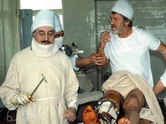
The method gained worldwide fame after Ilizarov treated the famous Italian explorer and traveller Carlo Mauri. Mauri had suffered a severe fracture of the tibia and had been treated in leading clinics around the world, without success. Only Ilizarov was able to help him. From then on, patients from all over the world, Serbia included, flocked to the little-known city of Kurgan in Siberia.

Today you no longer need to travel anywhere. Our team includes the Russian physician Professor Aleksandr Artemev, a world-class specialist and the author of the book “Reconstructive and Aesthetic Surgery of the Lower Limbs”.

Gavriil Ilizarov and Carlo Mauri in the operating room in Kurgan
Gavriil Ilizarov and the Italian explorer Carlo Mauri, Kurgan
  • 1 mmper day — the rate of gradual bone lengthening
  • ≈ 3months — average time in the frame for a deformity correction
  • 24 hafter surgery — weight-bearing on the operated leg is allowed
  • 2legs — can be operated on at the same time

What the frame makes possible

The frame is attached to the bone with thin wires through the skin, without large incisions and without plates or nails inside the bone. After an osteotomy through a puncture of a few millimetres, daily adjustment of the frame brings the bone gradually and almost painlessly into the desired position, and thanks to the stable fixation the patient bears weight on the leg from the first days. With the same frame, and without further incisions, the angle, displacement, rotation and length of the bone are corrected at the same time.

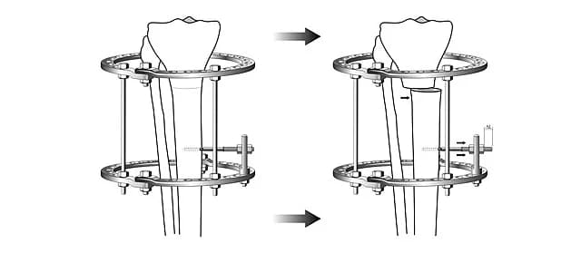
Angular correction
Angular correctionAfter the osteotomy the axis of the bone is gradually returned to the correct position — for any degree and type of curvature.
Medialisation
MedialisationThe lower bone fragment is gradually shifted inwards, improving the shape of the lower leg.
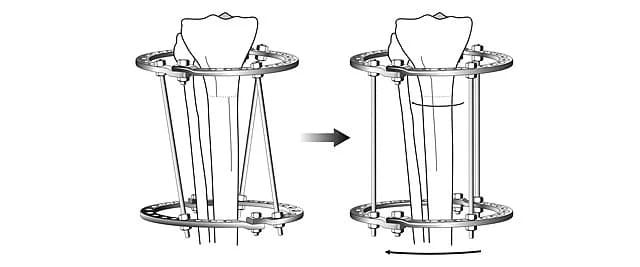
Rotational correction
Rotational correctionTorsion of the lower leg or thigh is corrected by rotating the rings of the frame, without new incisions.
Lengthening
LengtheningThe rings are moved apart about 1 mm a day and the gap between the fragments fills with new bone of the patient's own.
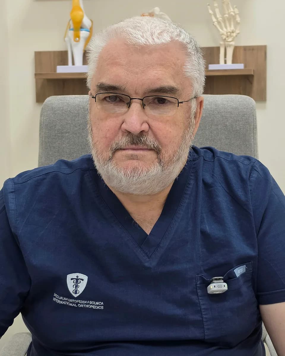
Prof. Dr. Aleksandr Artemev — Orthopedic and trauma surgeon, Doctor of Medical Sciences, Belgrade, Serbia

Consultant

Prof. Dr. Aleksandr Artemev

Orthopedic and trauma surgeon, Doctor of Medical Sciences

Orthopedic and trauma surgeon and Doctor of Medical Sciences, author of the book “Reconstructive and Aesthetic Surgery of the Lower Limbs”, of a classification of leg shapes (2001) and of a doctoral thesis on correcting leg shape and length in reconstructive and aesthetic surgery (2003). He has performed more than 1,500 leg correction and lengthening operations and personally follows every patient to the end of treatment.

Areas of expertise

What our clinic can do

The following list of Professor Aleksandr Artemev's main areas of expertise shows what our clinic can do:

  1. 01The mechanical axis of the lower limbs — what is normal
  2. 02General principles of deformity correction
  3. 03General principles of limb lengthening
  4. 04Knee osteotomy
  5. 05Correction of O-shaped knee deformity (bow legs)
  6. 06Correction of X-shaped knee deformity (knock knees)
  7. 07Visual correction of leg deformity
  8. 08Fracture treatment with the Ilizarov method
  9. 09Treatment of post-traumatic osteomyelitis
  10. 10Treatment of nonunion (pseudarthrosis)

In depth

More about the individual areas

Texts by Prof. Dr. Aleksandr Artemev and our team on the individual areas of treatment — from aesthetic correction to limb salvage after severe injuries.

03

General principles of limb lengthening

How bone is lengthened in the Ilizarov frame, at what rate, how long treatment takes and where the limits of safe lengthening lie.

All modern limb-lengthening operations around the world are based on Ilizarov's discovery, “The general biological property of tissues to respond to stretching with growth and regeneration — the Ilizarov effect” (1973). It means that if tissues are stretched gradually at a certain rate, new, identical tissue forms (regenerates).

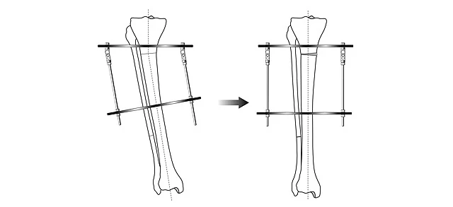
The lengthening technique is simple. After the osteotomy and application of the frame, the bone fragments are gradually pulled apart by increasing the distance between the proximal and distal supports on the threaded rods that connect them.
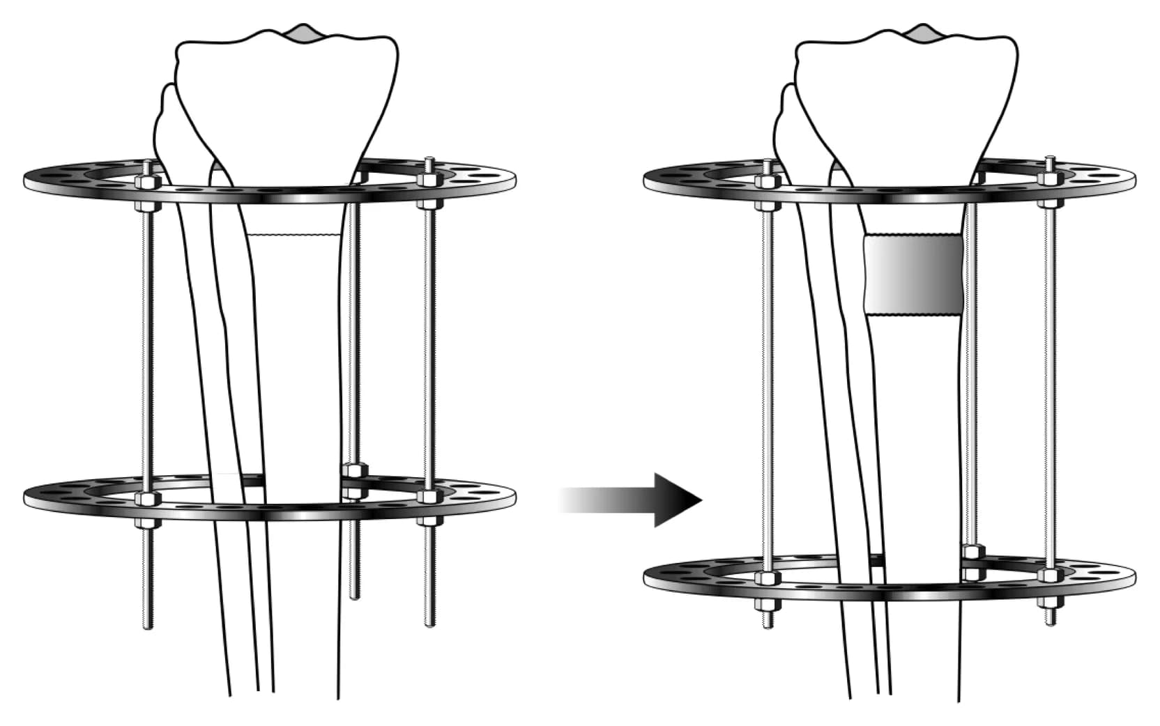
Diagram of tibial lengthening in the Ilizarov frame
Fig. 1. Diagram of lengthening (using the tibia as an example).

Numerous experimental and clinical studies have established the optimal distraction rate of 1 mm per day. The more even the distraction, the shorter the period of regenerate bone formation and the shorter the treatment.

In practice, patients carry out the distraction themselves by increasing the distance between the supports on the rods. The nuts are turned by 90° every 6 hours. The pitch of the M6 thread on the rods of the Ilizarov frame is 1 mm, so one full 360° turn of the nut per day equals 1 mm of lengthening. Distraction begins 5–7 days after the osteotomy.

The total time in the frame consists of a distraction period and a fixation period. During distraction, the distance between the fragments increases and the segment lengthens. During fixation, the regenerate bone matures and finally joins the bone fragments firmly together.
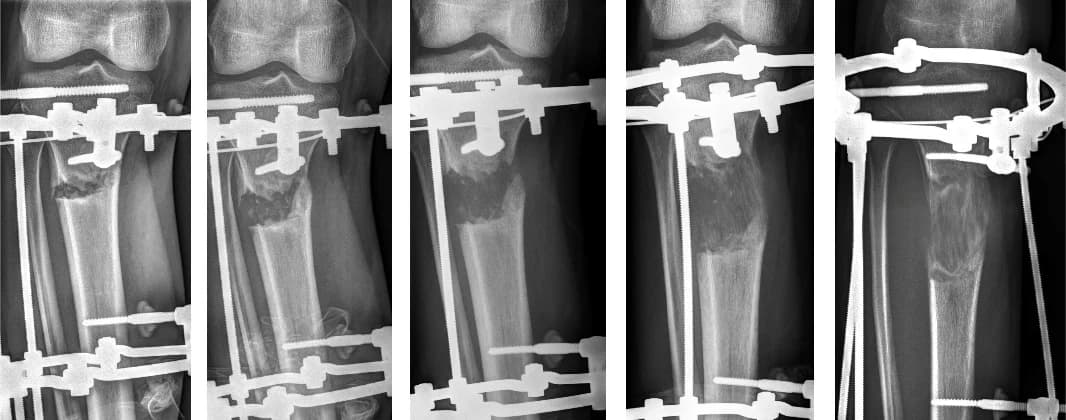
X-rays of the lower leg during distraction — weeks 1 to 12
Fig. 2. X-rays of the patient's right leg after 1, 3, 5, 9 and 12 weeks. The distraction process (from 0 to 7 cm) plus gradual correction of the axis.
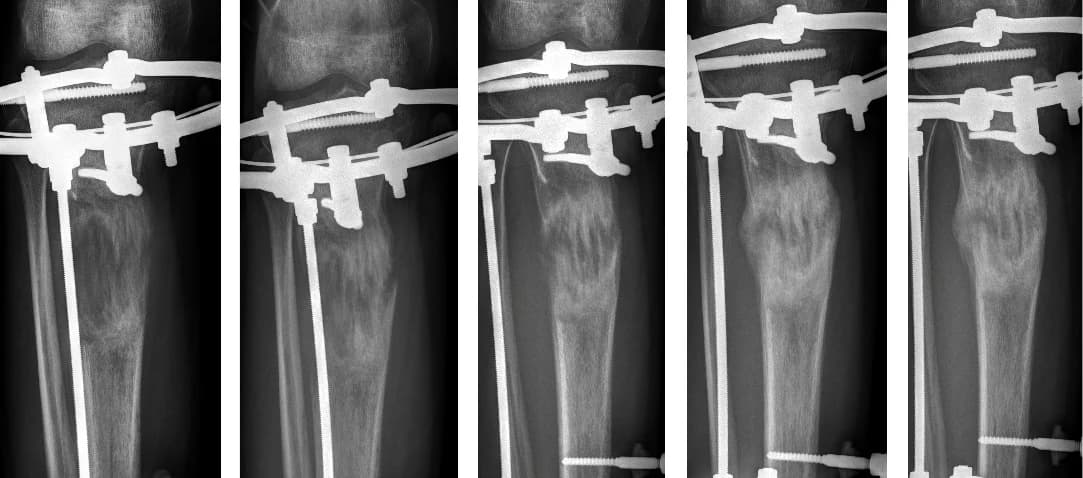
X-rays of the lower leg during fixation — weeks 12 to 27
Fig. 3. X-rays of the same patient 12, 16, 20, 24 and 27 weeks after surgery. The fixation phase is under way; the gradual remodelling and consolidation of the regenerate bone is visible.

Treatment takes a long time. Its approximate duration can be calculated with the formula:

T = 2.5 + Lwhere T is the treatment period in months and L the amount of lengthening in centimetres.

A 5 cm lengthening of the tibia will therefore take 7.5 months on average. It should be noted that these time frames are very approximate and that the duration of treatment cannot be predicted in every individual case.

It is important to remember that the body's resources have their limits. Lengthening a limb by large amounts is dangerous. We are used to expressing the amount of lengthening in absolute units of length, in centimetres, but it is more accurate to speak of the relative amount — a percentage of the original length. The optimal amount of lengthening is no more than 10–15% of the original length of the segment. For example, if the tibia is 35 cm long, the acceptable amount of lengthening is 53 mm (15% of 35 cm).

Two main factors limit lengthening:

  • the regenerate does not form (it dissolves and takes on an hourglass shape);
  • the tendons resist the lengthening.

The main recommendation is therefore not to be greedy and not to attempt to lengthen a limb by a large amount. That leads to serious complications (long treatment periods, joint contractures and subluxations). It is better to limit the lengthening to 10–15% of the original length.

If a significant lengthening is needed, it is best done in several stages. For example, if one leg is 10 cm shorter, it can be lengthened twice by 5 cm, with a recovery period between the two operations.

In reconstructive surgery of the lower limbs, lengthening is used in two situations:

  • to correct a leg length discrepancy (lengthening the shorter limb);
  • to increase height in certain conditions (for example achondroplasia) or at the patient's request. In such cases both legs are usually lengthened at the same time.
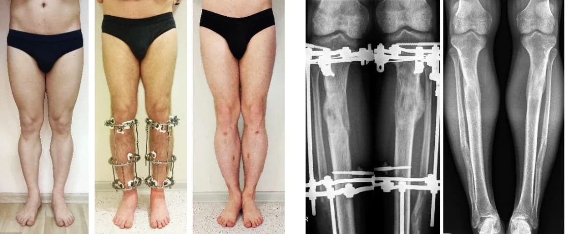
Patient before, during and after bilateral 7 cm lower-leg lengthening, with X-rays
Fig. 4. Left: a patient whose legs were lengthened by 7 cm (before, during and after treatment); right: X-rays during and after treatment.
Patient before and after 4 cm lengthening of the right tibia, with X-rays
Fig. 5. Left: a patient whose right tibia was lengthened by 4 cm (before and after treatment); right: X-rays before and after.

The regenerate bone formed during distraction and subsequent fixation is truly the patient's own bone. A few years after surgery the site of the osteotomy and lengthening is practically impossible to find on X-rays. The bone in this area is as strong as it was before the operation.

Lengthening procedures, including the operation and the postoperative period, require a responsible and serious attitude on the part of the patient. Regular communication with the treating doctor is essential.

The details of the forthcoming treatment, the prospects and the prognosis for achieving the desired result are discussed during the consultation with Professor Aleksandr Artemev.
04

Knee osteotomy

Unloading the damaged part of the knee by correcting the leg axis — an alternative to knee replacement in the early and middle stages of osteoarthritis.

When the leg axis is shifted into varus or valgus, one compartment of the knee carries almost the entire load and the cartilage on that side wears rapidly. A corrective osteotomy of the thigh or lower leg restores the mechanical axis and redistributes the load: the damaged compartment is unloaded by about 70%, pain decreases and the remaining cartilage gets a chance to recover.

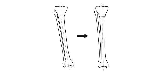
Diagram of tibial axis correction by osteotomy
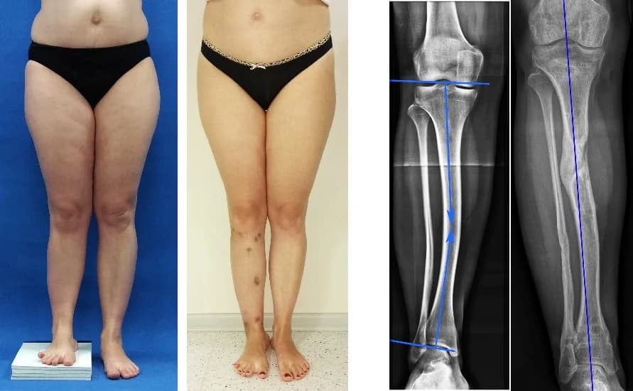
The osteotomy restores the mechanical axis and unloads the damaged compartment of the knee.

Osteotomy is considered the gold standard for grade 1 to 3 knee osteoarthritis in patients aged 15 to 70 — performed in time, it postpones or entirely removes the need for a knee replacement in many patients. The basic investigation is a standing full-length X-ray of both legs, which shows the size of the deformity, the affected compartment and the optimal level for the osteotomy.

Compared with plate fixation, the Ilizarov frame allows both legs to be operated on at once, the axis to be adjusted precisely and gradually — including deformities in several planes — and the patient to walk with weight-bearing from the first days. Where indicated, the osteotomy is combined with knee arthroscopy in the same session, and a slight lengthening of the bone restores tension in the collateral ligaments and further stabilises the joint.

05

Correction of O-shaped knee deformity (bow legs)

Correction of varus deformity of the lower leg and thigh — the most common reason for aesthetic leg correction, and prevention of knee osteoarthritis.

True bow legs (varus deformity) are recognised by the knees not touching when the feet are together, leaving a gap along the inner contour of the legs from the groin to the feet. They must be distinguished from so-called false curvature, where the bones are straight and the impression of bowed legs is created by the distribution of the soft tissues of the lower leg. In Prof. Artemev's classification of 2001 it is precisely this distinction that determines the method of correction. Bow legs are by far the most common reason for aesthetic correction — 15 to 20 times more common than knock knees.

A deviation of the leg's mechanical axis is not merely an aesthetic problem: it overloads the inner compartment of the knee, so the cartilage wears faster and knee osteoarthritis appears earlier. Correction is therefore recommended as soon as possible after growth is complete. True curvature cannot be corrected with exercise, massage or braces — a deformed bone cannot be stretched straight. In children the deformity is usually observed until about the age of 8; if a disease is the cause (for example Blount's disease) surgery is done earlier, otherwise one waits for the growth plates to close, between the ages of 14 and 18.

The correction is performed through an osteotomy made via a puncture of about 5 mm, most often on the lower leg, which is technically simpler and safer than the thigh. In the Ilizarov frame the bone is then gradually brought into the correct axis, and where needed the rotation, the sideways shift of the lower fragment (medialisation, added at the patient's request in about 60% of cases), the position of the fibular head and the length of the leg are corrected at the same time — most often a lengthening of 2 to 4 cm for better proportions. The patient takes part in deciding the final shape of the legs, and the frame, weighing about 900 g, can be hidden under loose clothing.

The frame is worn for about three months on average; each centimetre of lengthening adds roughly a month. In suitable cases so-called express methods are possible, in which the frame is combined with an intramedullary nail, so it is worn for a much shorter time and active rehabilitation begins after two to three weeks. Which method is most suitable is decided at the consultation on the basis of the examination and a standing full-length X-ray of both legs.

06

Correction of X-shaped knee deformity (knock knees)

Correction of valgus deformity — a rarer but often more pronounced form of leg curvature, which can progress with age.

True knock knees (valgus deformity) are recognised by the gap between the feet when the knees are together in a relaxed stance. An axis deviation of up to 10–15° is considered a cosmetic problem, more than 15° an orthopedic one. This leg shape is an ethnic trait, more common among Europeans; surgical correction of knock knees is sought 10 to 15 times less often than for bow legs, and men present two to three times more often than women.

In trousers, legs with a slight deformity look straight, while with a pronounced deformity clothes fit badly — a frequent reason for seeing a doctor. Valgus deformity is often accompanied by underdevelopment of the femoral condyles and flat feet, and it can progress with age, so correction is recommended as early as possible. Static deformities — valgus knees, bunions (hallux valgus), flat feet — are linked to the way connective tissue develops: when one is found, examination for the others is advisable, and children suspected of a deformity should see an orthopedic surgeon at least once a year.

The principle of surgical treatment is the same as for bow legs, only the shift is directed the opposite way. Three options are available: classic correction in the Ilizarov frame, the express method, and the improved express method with intramedullary nail fixation, in which active rehabilitation begins around day 19 after surgery. The legs can be lengthened at the same time for a more harmonious appearance. It is the curved segment itself that is corrected; correction of the lower leg is technically simpler and causes fewer complications than operations on the thigh.

08

Fracture treatment with the Ilizarov method

Stabilisation and healing of complex, open, comminuted and periarticular fractures without internal implants — even when other methods are not possible.

In open and severely comminuted fractures, fractures with soft-tissue damage, gunshot and infected fractures, inserting plates or nails carries a high risk of infection and nonunion. The Ilizarov frame fixes the bone with thin wires away from the fracture site, without foreign material in the wound, so the soft tissues and wounds can be treated while the bone heals.

The fragments can be repositioned precisely and gradually in the frame even after surgery, without reopening, which is especially important in complex fractures near joints — ankle and pilon fractures, fractures of the elbow and wrist. The stability of the frame allows early mobilisation of the neighbouring joints and weight-bearing, preserving function and preventing contractures.

The method is also the answer for fractures that have healed in the wrong position (malunion): the bone is cut again at the site of the deformity and gradually straightened in the frame, with any shortening made up. The frame is worn until the bone has fully united — usually several months for complex fractures of the lower leg — and in the final phase of treatment the patient as a rule walks without crutches.

09

Treatment of post-traumatic osteomyelitis

Curing chronic bone infection after fractures or surgery — by radical removal of the diseased tissue and replacement of the bone defect with the Ilizarov method.

Osteomyelitis is a purulent infection of bone that most often develops after open fractures, after surgery with plates, nails or joint prostheses, and around the wires of external fixators. Bacteria are a necessary but not a sufficient condition: what matters most is the poor state of the bone and surrounding soft tissues — impaired circulation and necrosis after severe injuries — and the presence of foreign material on which the infection persists. Antibiotics are an indispensable part of treatment, but on their own they cannot cure chronic bone infection.

The basis of treatment is surgical, and in a defined order: removal of foreign material, radical resection of non-viable, infected bone and soft tissue, then achieving union and load-bearing capacity of the segment, and finally reconstruction — restoring length, treating joint contractures and covering soft-tissue defects.

The Ilizarov method is ideal for this because it stabilises the bone without any implant in the infected area and replaces the resulting bone defect with the patient's own bone — by so-called bone transport (bifocal osteosynthesis) or by acute shortening followed by lengthening. Where needed it is combined with vacuum-assisted wound therapy (VAC), an antibiotic-cement-coated intramedullary nail or transfer of the fibula into a tibial defect (fibula pro tibia). Treatment is staged and takes months, but it allows even long-standing osteomyelitis to be permanently cured while preserving the limb.

10

Treatment of nonunion (pseudarthrosis)

Treatment of un-united fractures and bone defects — by compression and stimulation of the bone's own regeneration, without bone grafts from other parts of the body.

We speak of delayed union when a fracture has not healed within the average time expected for that bone, and of nonunion (a “false joint”) when it has not healed after twice that period. A fracture of the femoral shaft, for example, usually heals in 3–4 months; if it has not united after 8 months, a nonunion has formed. Bone defects after injuries, surgery or tumours are a particular problem: the fragments are not in contact at all, so healing without surgery is impossible.

A distinction is made between hypertrophic (stiff) nonunions, with little mobility and a partly load-bearing limb — for which several months of stable fixation are sometimes sufficient — and hypotrophic (mobile) nonunions with marked instability, which require surgery at the site of the nonunion: freshening of the bone ends, firm contact between them and reliable fixation.

The Ilizarov frame provides exactly that: measured compression of the fragments that promotes union, alternating compression and distraction that activates regeneration, and at the same time correction of deformity and replacement of shortening or defect with the patient's own bone. This avoids the most common mistake — grafting bone from the pelvis or fibula, which often fails to achieve union and carries the risk of infection and even larger defects. The result is a united, load-bearing and correctly aligned bone.

Treatment pathway

What treatment looks like — step by step

  1. 01

    Consultation

    Examination or remote assessment based on photographs and X-rays; a conversation about goals, options, duration and cost of treatment.

  2. 02

    Preparation

    A standing full-length X-ray of both legs, CT where needed, laboratory tests and a physician's check-up — all carried out at our hospital.

  3. 03

    Surgery

    A minimally invasive osteotomy through a puncture of a few millimetres and application of the Ilizarov frame — on both legs at once where appropriate.

  4. 04

    Correction and distraction

    From day 5 to 7, gradual adjustment of the frame according to plan — about 1 mm a day — with weight-bearing walking and regular check-ups.

  5. 05

    Consolidation

    The new bone matures and hardens; loading is gradually increased to full weight-bearing without aids.

  6. 06

    Frame removal and rehabilitation

    A short procedure under brief anaesthesia, followed by physiotherapy and a return to everyday activities.

For international patients

Remote assessment — before you travel

Many of our patients come from abroad. So that we can give you, before you arrive, an accurate assessment of the options, duration and cost of treatment for your case, please e-mail us:

We will reply with an assessment, an exact price and the doctor's comments.

office@trifunovic-ortopedija.rs

  1. 1Your name, gender and age
  2. 2Height and weight (approximate figures are fine) and your country of residence
  3. 3Photographs of your legs — standing relaxed, from the waist to the feet, taken from knee height at a distance of 2.5–3 m
  4. 4X-rays, if you have them
  5. 5For leg lengthening: by how many centimetres you would like to lengthen, thighs or lower legs, and which method you would prefer

Guidance

Before your consultation and surgery

01

The scope of examinations required for surgery at our clinic

Laboratory tests no older than 10 days: blood count and urinalysis; biochemistry (glucose, bilirubin, creatinine, urea, ALT, AST); coagulation profile; blood group and Rh factor; tests for HIV, hepatitis B and C and syphilis.

Instrumental examinations: a chest X-ray no older than one year; an ECG with a physician's check-up (a paediatrician's for children); an ultrasound of the leg vessels where indicated; an X-ray or CT of the limbs.

If you are in good health you do not need to do any of this in advance — all examinations are done at our hospital and their cost is included in the price of treatment. If you have chronic conditions, have the necessary check-ups where you live to rule out contraindications for surgery and send us the results.

02

What the X-ray examination should include

For leg-shape correction, knee osteotomy and lengthening, a standing full-length X-ray of both legs is required — from the hips to the ankles, both legs on one image, with the kneecaps facing straight forward. Only such an image shows the mechanical axis of the leg and allows the correction to be planned precisely.

For fractures, nonunions and osteomyelitis, an X-ray of the affected segment in two views (front and side) is required, and a CT scan where needed. Digital images (DICOM files on a disc or USB stick) are always better than a photograph of an X-ray; if you do not have them, see the guidance below on photographing an X-ray.

03

How to take photos (and video) for an online consultation

Stand relaxed, without tensing. The legs should barely touch: at the feet for bow legs, at the knees for knock knees. Use the flash and a flat, light, matte background (a door or a wall); avoid mirrors, backlighting and glossy surfaces, which cause glare and distort the outline of the legs.

Selfies are not recommended — have someone else take the photo or use a timer. The camera should be at knee height, parallel to the axis of the legs, 2.5–3 m away (the person taking the photo should crouch). The frame should include the body from the navel to the toes; for leg lengthening also send a full-length photo taken from waist height. You may cover or blur your face.

If you also send a video, film a few steps towards and away from the camera, from the same distance and under the same conditions.

04

How to photograph an X-ray taken on film

Turn off the flash. Place the film on a light box, or hold it against a monitor showing a blank white page full-screen — that gives an even, bright background. Do not photograph the X-ray against a window.

Hold the camera exactly parallel to the film so that the whole image is in the frame and free of reflections. The same applies when photographing an X-ray from a computer screen. If you have the images in digital form (CD, USB, DICOM file), send those instead of a photograph.

Prices

Cost of treatment with the Ilizarov method

Full price list →

Prices are base rates in Serbian dinars (RSD), by category of complexity. The exact price for your case is given after the doctor's assessment.

Price List of Services by Prof. Dr. Artemev — Treatment with the Ilizarov Method

  • Category 1 — osteotomy or uncomplicated fracture of one segment400,000.00 RSD
  • Category 2 — osteotomy of two segments, lengthening of one segment or a complex fracture700,000.00 RSD
  • Category 3 — complex correction of two segments, lengthening of two segments or a complex fracture1,200,000.00 RSD
  • Category 4 — complex correction of two segments with lengthening, bone defect, nonunion, osteomyelitis or multi-stage treatment2,000,000.00 RSD

FAQ

Questions patients ask us most often

Didn't find your answer? Book a consultation or write to us — we also answer questions sent by e-mail from abroad.

The price list is available on our website — the prices shown are base rates by category of complexity. For an accurate estimate of the cost of surgery and the entire course of treatment in your specific case, send your name, gender, age, height and weight, country of residence and photographs of your legs to office@trifunovic-ortopedija.rs; for lengthening, also state by how many centimetres, thighs or lower legs, and which method you would prefer. We will reply with an exact price and the doctor's comments.

As a rule these procedures are performed after skeletal growth and development are complete — that is, after the age of 18. If a specialised X-ray shows that the growth plates have closed, they can be performed earlier. For congenital or acquired deformities in children the optimal age for surgery is determined individually by the doctor, according to the underlying diagnosis and the severity of the deformity — send us photographs of the affected area and X-rays, if available, and the doctor will reply.

You need laboratory tests no older than 10 days (blood count, urinalysis, biochemistry, coagulation profile, blood group, and tests for HIV, hepatitis B and C and syphilis), an ECG with a physician's check-up (a paediatrician's for children), an X-ray or CT of the limbs and, where indicated, an ultrasound of the leg vessels. If you are in good health you do not need to do any of this in advance — everything is done at our hospital and the cost of the examinations is included in the price of treatment. If you have chronic conditions, have the necessary check-ups where you live to rule out contraindications and send us the results.

For a deformity correction the frame is worn for about three months on average; for lengthening, allow roughly 2.5 months plus one month per centimetre. Express methods with an intramedullary nail shorten the time in the frame to a few weeks, or to the distraction period only. Thanks to the stable fixation you bear weight on the leg the day after surgery, at first with walking aids, and in the final phase of treatment most patients walk without crutches. Removing the frame too early or overloading the leg can deform the still immature new bone, so following the instructions is essential.

The operation is performed under anaesthesia, and the gradual adjustment of the frame, which the patient carries out according to instructions, is practically painless. A standard lower-leg frame weighs a little over 900 grams and can be hidden under loose clothing. Showering is allowed and recommended — the frame is protected with a plastic bag. Staff train you in caring for the wire sites; in case of any redness, swelling or pain you should contact the doctor at once.

Removal takes a few minutes and is done under brief general anaesthesia; when both legs have been operated on, the frames are removed one at a time, one to two weeks apart. The wire sites leave small scars that mature over a year — during that time they should be protected from the sun. If they still bother you after a year, they can be treated with a laser or removed surgically.

Exercise, massage, straps and various stretching devices cannot correct true curvature — a deformed bone cannot be straightened by stretching. Without an external frame, correction is possible with a plate or an intramedullary nail, with certain limitations: it is harder to lengthen the leg or shift the lower fragment at the same time, and the patient cannot take part in judging the shape during correction. Which method is best for you is assessed at the consultation.

24 / 7

Book an examination and consultation

Your health is our priority. Make an appointment with our expert doctors today and take the first step towards better health. It's quick and easy!